How is Centronuclear Myopathy diagnosed?
See how Centronuclear Myopathy is diagnosed. Which specialists are essential to meet, what tests are needed and other useful information for the diagnosis of Centronuclear Myopathy
Centronuclear Myopathy (CNM) is a rare genetic disorder that affects the muscles, causing weakness and potential complications in movement. Diagnosing CNM involves a comprehensive evaluation of the patient's medical history, physical examination, and a series of specialized tests.
Medical History: The first step in diagnosing CNM is to gather a detailed medical history of the patient. This includes information about the onset and progression of symptoms, family history of muscle disorders, and any other relevant medical conditions. Understanding the patient's background helps the healthcare provider to identify potential risk factors and genetic patterns associated with CNM.
Physical Examination: A thorough physical examination is conducted to assess muscle strength, tone, and reflexes. The healthcare provider will look for specific signs such as muscle weakness, reduced muscle bulk, and abnormal muscle tone. They may also evaluate other body systems to rule out alternative causes for the symptoms.
Genetic Testing: Genetic testing plays a crucial role in diagnosing CNM. It involves analyzing the patient's DNA to identify specific genetic mutations associated with the condition. The most common genes linked to CNM are MTM1, DNM2, and BIN1. Genetic testing can be performed using various techniques, including targeted gene sequencing, whole-exome sequencing, or even whole-genome sequencing. These tests help confirm the presence of genetic mutations that cause CNM.
Muscle Biopsy: A muscle biopsy is often recommended to support the diagnosis of CNM. During this procedure, a small sample of muscle tissue is extracted, typically from the thigh or upper arm. The sample is then examined under a microscope to assess the structure and appearance of muscle fibers. In CNM, muscle biopsy findings often reveal centrally located nuclei within the muscle fibers, which is a characteristic feature of the condition.
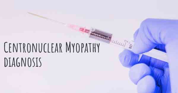
Electromyography (EMG): EMG is a specialized test that measures the electrical activity of muscles. It involves inserting a fine needle electrode into the muscle to record the muscle's response to electrical stimulation. In CNM, EMG may show abnormal electrical patterns, indicating muscle dysfunction.
Muscle Imaging: Imaging techniques such as magnetic resonance imaging (MRI) or computed tomography (CT) scans can provide valuable information about muscle structure and any associated abnormalities. These imaging studies help visualize muscle size, fat infiltration, and potential structural changes that may be indicative of CNM.
Other Tests: Additional tests may be conducted to rule out other muscle disorders or to assess the severity and potential complications of CNM. These may include blood tests to measure muscle enzyme levels, nerve conduction studies to evaluate nerve function, or cardiac evaluations to assess heart involvement.
It is important to note that the diagnostic process for CNM may vary depending on the individual case and the expertise of the healthcare provider. Consulting with a medical professional experienced in neuromuscular disorders is crucial for an accurate diagnosis and appropriate management of CNM.
Posted Feb 18, 2018 by Kimberly 1850