How is Graves disease diagnosed?
See how Graves disease is diagnosed. Which specialists are essential to meet, what tests are needed and other useful information for the diagnosis of Graves disease
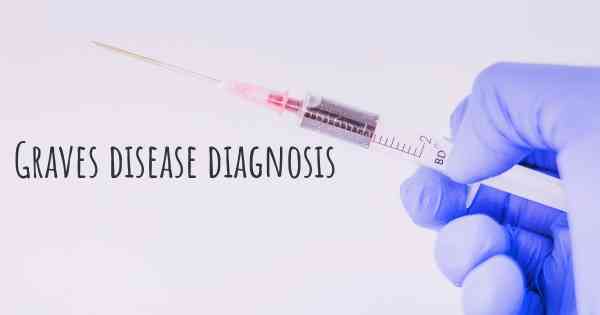
Diagnosing Graves' Disease
Graves' disease is an autoimmune disorder that affects the thyroid gland, leading to the overproduction of thyroid hormones. It is important to diagnose this condition promptly to initiate appropriate treatment and prevent potential complications. Diagnosing Graves' disease involves a combination of medical history assessment, physical examination, and various diagnostic tests.
Medical History Assessment
The first step in diagnosing Graves' disease is a comprehensive medical history assessment. The healthcare provider will inquire about the patient's symptoms, such as unexplained weight loss, rapid heartbeat, anxiety, tremors, heat intolerance, and changes in bowel movements. They will also ask about any family history of thyroid disorders or autoimmune diseases, as these can increase the risk of developing Graves' disease.
Physical Examination
During the physical examination, the healthcare provider will look for physical signs associated with Graves' disease. These may include bulging eyes (exophthalmos), swelling or enlargement of the thyroid gland (goiter), rapid heartbeat (tachycardia), and tremors in the hands. The presence of these signs, along with the patient's symptoms, can provide valuable clues for the diagnosis.
Thyroid Function Tests
Thyroid function tests are crucial in diagnosing Graves' disease. These tests measure the levels of thyroid hormones (T3 and T4) and thyroid-stimulating hormone (TSH) in the blood. In Graves' disease, the levels of T3 and T4 are typically elevated, while TSH levels are suppressed. These findings indicate an overactive thyroid gland.
Thyroid Antibody Tests
Another important diagnostic tool is thyroid antibody testing. In Graves' disease, the body produces specific antibodies called thyroid-stimulating immunoglobulins (TSIs) or thyroid receptor antibodies (TRAb). These antibodies stimulate the thyroid gland to produce excess hormones. Measuring the levels of TSIs or TRAb in the blood can help confirm the diagnosis of Graves' disease.
Radioactive Iodine Uptake (RAIU) Test
The radioactive iodine uptake (RAIU) test is often performed to assess the function of the thyroid gland. In this test, a small amount of radioactive iodine is administered orally, and its uptake by the thyroid gland is measured. In Graves' disease, the thyroid gland typically takes up an excessive amount of radioactive iodine, indicating increased thyroid activity.
Thyroid Ultrasound
A thyroid ultrasound may be recommended to evaluate the size and structure of the thyroid gland. This imaging test uses sound waves to create images of the thyroid. It can help identify any abnormalities, such as nodules or enlargement, which may be present in Graves' disease.
Eye Examination
Since Graves' disease can cause eye-related symptoms, an eye examination by an ophthalmologist is often necessary. The ophthalmologist will assess the patient's eye movements, visual acuity, and look for signs of eye involvement, such as bulging eyes, redness, or swelling. They may also perform additional tests, such as visual field testing or orbital imaging, to evaluate the extent of eye complications.
Other Tests
In some cases, additional tests may be required to rule out other potential causes or assess the overall health of the patient. These may include blood tests to evaluate liver and kidney function, as well as an electrocardiogram (ECG) to assess heart rhythm.
Consultation with an Endocrinologist
Given the complexity of Graves' disease, a consultation with an endocrinologist, who specializes in hormonal disorders, is often recommended. The endocrinologist will review the patient's medical history, physical examination findings, and the results of various tests to confirm the diagnosis and develop an appropriate treatment plan.
Conclusion
Diagnosing Graves' disease involves a comprehensive approach that includes medical history assessment, physical examination, and various diagnostic tests. Thyroid function tests, thyroid antibody tests, radioactive iodine uptake (RAIU) test, thyroid ultrasound, and eye examination are among the key tools used to diagnose this autoimmune disorder. Consulting with an endocrinologist is crucial to confirm the diagnosis and initiate appropriate treatment.
Posted Mar 17, 2017 by Abby 200
Posted Mar 17, 2017 by Emma 3770