How is Myasthenia Gravis diagnosed?
See how Myasthenia Gravis is diagnosed. Which specialists are essential to meet, what tests are needed and other useful information for the diagnosis of Myasthenia Gravis
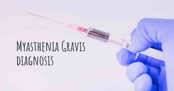
Diagnosing Myasthenia Gravis
Myasthenia Gravis (MG) is a chronic autoimmune neuromuscular disorder that affects the communication between nerves and muscles. It leads to muscle weakness and fatigue, particularly in the voluntary muscles. Diagnosing MG can be challenging as its symptoms can be similar to other conditions. However, there are several key diagnostic methods that healthcare professionals use to identify and confirm the presence of Myasthenia Gravis.
Clinical Evaluation and Medical History
The initial step in diagnosing Myasthenia Gravis involves a comprehensive clinical evaluation and a detailed medical history. The healthcare provider will discuss the patient's symptoms, their onset, and their progression. They will also inquire about any family history of autoimmune disorders, as MG can have a genetic component. This information helps the doctor understand the patient's condition better and determine the appropriate diagnostic tests.
Physical Examination
A thorough physical examination is crucial in diagnosing Myasthenia Gravis. The healthcare provider will assess the patient's muscle strength, reflexes, and coordination. They will look for specific signs of muscle weakness, such as drooping eyelids (ptosis), difficulty swallowing (dysphagia), and impaired speech. These physical manifestations, combined with the patient's medical history, can provide valuable clues for further investigation.
Neurological Examination
Since Myasthenia Gravis affects the neuromuscular junction, a neurological examination is an essential part of the diagnostic process. The healthcare provider will evaluate the patient's cranial nerves, muscle tone, and muscle strength. They may perform tests like the ice pack test, where an ice pack is applied to a weak muscle to observe if it improves temporarily. These tests help differentiate MG from other neuromuscular disorders.
Electrodiagnostic Tests
Electrodiagnostic tests are commonly used to diagnose Myasthenia Gravis. The most frequently employed tests include:
- Repetitive Nerve Stimulation (RNS): This test involves delivering repetitive electrical stimuli to a nerve while measuring the muscle response. In MG, the muscle response progressively weakens with repeated stimulation.
- Single Fiber Electromyography (SFEMG): SFEMG measures the electrical activity of individual muscle fibers. In Myasthenia Gravis, there is an increased jitter (variation in the time it takes for muscle fibers to respond) due to the disrupted neuromuscular transmission.
Blood Tests
Blood tests are an essential component of the diagnostic process for Myasthenia Gravis. The presence of specific autoantibodies can help confirm the diagnosis. The two primary autoantibodies associated with MG are:
- Acetylcholine Receptor Antibodies (AChR): Approximately 85% of MG patients have AChR antibodies. Blood tests can detect these antibodies, which bind to acetylcholine receptors and impair neuromuscular transmission.
- Muscle-Specific Kinase Antibodies (MuSK): Around 5-10% of MG patients have MuSK antibodies. These antibodies target a protein involved in the functioning of the neuromuscular junction.
Imaging Studies
Imaging studies are not typically used for diagnosing Myasthenia Gravis but may be performed in certain cases. Chest CT scans can help identify thymomas (tumors of the thymus gland) that are associated with MG. Thymomas can contribute to the development or exacerbation of MG symptoms.
Tensilon Test
The Tensilon test, also known as the edrophonium test, is a diagnostic tool used to confirm the presence of Myasthenia Gravis. During this test, a small amount of a medication called edrophonium chloride (Tensilon) is injected intravenously. If the patient has MG, there is a temporary improvement in muscle strength within a few minutes of the injection. This test helps differentiate MG from other conditions that cause muscle weakness.
Consultation with Specialists
Diagnosing Myasthenia Gravis often involves collaboration between different healthcare professionals. Neurologists, rheumatologists, and other specialists with expertise in neuromuscular disorders may be consulted to confirm the diagnosis and develop an appropriate treatment plan.
It is important to note that the diagnostic process for Myasthenia Gravis can vary from person to person. Some individuals may have clear clinical symptoms and positive autoantibodies, making the diagnosis relatively straightforward. However, in other cases, additional tests and consultations may be required to reach a definitive diagnosis.
Tension test, AChr and Ab test.
Posted Mar 12, 2017 by Ara Eliana 1145
Single Fiber EMG
Exam
Posted May 24, 2017 by Sherri 720
Posted May 24, 2017 by Gary 1500
MRI of my brain was taken first. The result was normal. Then an EMG examination was performed. EMG examined my face. These values were compatible with the disease. Then I went to the lung tomography. The thymus gland seemed irritating. In this process, the doctor makes the necessary explanation and diagnosis. Six months later, I'm going to the hospital again for lung tomography. I'm using Mestinon right now.
The doctor of neurology is very important. Do not interrupt your controls. surely go.
Posted Dec 21, 2018 by Damla 2100
Laboratories blood
RX of thorax (to know if you have a tumor)
Examination to know which type of antibodies you have
Test Jolly or stimulation repetitive
Electromyography single fiber
Exam general doctor, in order to measure the bodily weakness
Posted Mar 14, 2017 by Alejandra 970
Posted May 24, 2017 by Meire Stella 1000
Posted May 24, 2017 by lorrayne 500
Search of acetylcholine
And a good neurologist knowledgeable in this pathology
Posted May 24, 2017 by Lilian Silva Santos 1000
Posted Sep 3, 2017 by Marife Loyola 650
Dai passes for eletroneuromiografia of 4 members (sup and inf), and can be facial also. Although giving multiple interference in this exsmes what feicha the diagnosis is the exsme blood antereceptor of aceltilcolina.
Posted Sep 13, 2017 by Paulo de Tarso c carvalho 1500
Posted Sep 13, 2017 by Lua 3536
Posted Sep 13, 2017 by Renata 4550
Posted Sep 13, 2017 by Monique 1500
Posted Sep 30, 2017 by Marcela 900