How is Pigmented villonodular synovitis diagnosed?
See how Pigmented villonodular synovitis is diagnosed. Which specialists are essential to meet, what tests are needed and other useful information for the diagnosis of Pigmented villonodular synovitis
Diagnosis of Pigmented Villonodular Synovitis (PVNS)
Pigmented villonodular synovitis (PVNS) is a rare benign condition that affects the synovial lining of joints, tendon sheaths, and bursae. It is characterized by the abnormal growth of the synovial tissue, leading to the formation of nodules and the accumulation of hemosiderin pigment. PVNS commonly affects the knee joint but can also occur in other joints such as the hip, ankle, and shoulder.
Medical History and Physical Examination
The diagnosis of PVNS begins with a thorough medical history and physical examination. The doctor will ask about the patient's symptoms, such as joint pain, swelling, stiffness, and limited range of motion. They will also inquire about any previous injuries or surgeries involving the affected joint.
During the physical examination, the doctor will carefully assess the affected joint, looking for signs of swelling, tenderness, and warmth. They will also evaluate the joint's range of motion and stability. These findings, combined with the patient's symptoms, can provide important clues to the presence of PVNS.
Imaging Studies
Imaging studies play a crucial role in the diagnosis of PVNS. The most commonly used imaging modalities include:
- X-rays: X-rays can help identify any bony changes associated with PVNS, such as erosion or cyst formation. However, they may not show the soft tissue abnormalities characteristic of PVNS.
- Magnetic Resonance Imaging (MRI): MRI is the imaging modality of choice for diagnosing PVNS. It provides detailed images of the soft tissues, allowing for the visualization of the abnormal synovial tissue, nodules, and joint effusion. MRI can also help determine the extent of the disease and its impact on surrounding structures.
- Ultrasound: Ultrasound can be used to evaluate joint effusion, synovial thickening, and the presence of nodules. It is a cost-effective and readily available imaging modality, but it may not provide as detailed information as MRI.
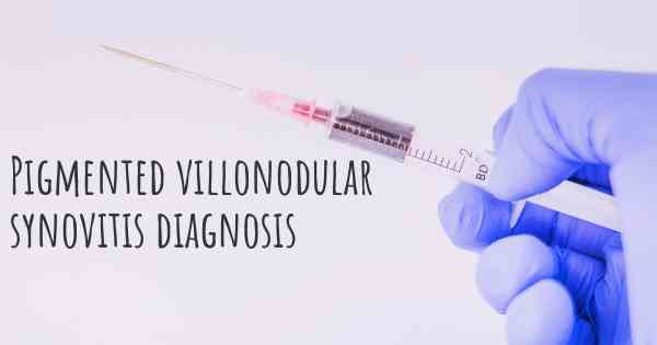
Biopsy
A biopsy is often necessary to confirm the diagnosis of PVNS. It involves the removal of a small sample of the synovial tissue for microscopic examination. There are two main types of biopsies:
- Needle Biopsy: A needle biopsy is a minimally invasive procedure where a thin needle is inserted into the affected joint under the guidance of imaging techniques. The needle is used to obtain a small tissue sample, which is then sent to the laboratory for analysis.
- Arthroscopic Biopsy: An arthroscopic biopsy is a more invasive procedure that requires a small incision to insert an arthroscope into the joint. The arthroscope is equipped with a camera and surgical instruments, allowing the surgeon to visualize and obtain a larger tissue sample for analysis.
The biopsy sample is examined by a pathologist who will look for the characteristic features of PVNS, including the presence of hemosiderin-laden macrophages, giant cells, and fibrous tissue proliferation.
Differential Diagnosis
Several other conditions can mimic the symptoms of PVNS, making the differential diagnosis important. Some of the conditions that need to be considered include:
- Osteoarthritis: Osteoarthritis can cause joint pain, swelling, and stiffness, similar to PVNS. However, PVNS typically affects younger individuals, while osteoarthritis is more common in older adults.
- Rheumatoid Arthritis: Rheumatoid arthritis can also present with joint swelling and stiffness. However, it usually affects multiple joints symmetrically, whereas PVNS is typically localized to a single joint.
- Gout: Gout is a form of arthritis caused by the deposition of uric acid crystals in the joints. It can cause sudden episodes of joint pain and swelling, which can be mistaken for PVNS.
Conclusion
Diagnosing pigmented villonodular synovitis (PVNS) requires a combination of medical history, physical examination, and imaging studies. X-rays, MRI, and ultrasound are commonly used to visualize the affected joint and assess the extent of the disease. However, a definitive diagnosis is often made through a biopsy, either by needle or arthroscopic biopsy. The biopsy sample is examined by a pathologist to confirm the presence of PVNS. Differential diagnosis is crucial to rule out other conditions that may present with similar symptoms. Early and accurate diagnosis of PVNS is essential for appropriate management and treatment planning.
Biopsy
Posted Jun 1, 2017 by ronan 200