How is Langerhans Cell Histiocytosis diagnosed?
See how Langerhans Cell Histiocytosis is diagnosed. Which specialists are essential to meet, what tests are needed and other useful information for the diagnosis of Langerhans Cell Histiocytosis
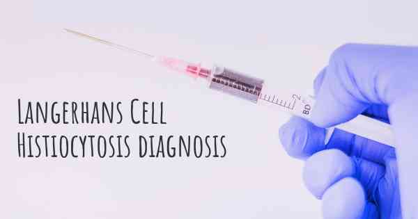
Diagnosis of Langerhans Cell Histiocytosis
Langerhans Cell Histiocytosis (LCH) is a rare disorder characterized by the abnormal accumulation and proliferation of Langerhans cells, a type of white blood cell, in various tissues and organs of the body. The diagnosis of LCH can be challenging due to its diverse clinical manifestations and the need to differentiate it from other similar conditions. However, a combination of clinical, radiological, histopathological, and immunohistochemical findings can help in establishing an accurate diagnosis.
Clinical Evaluation
The initial step in diagnosing LCH involves a thorough clinical evaluation, which includes a detailed medical history and physical examination. The healthcare provider will inquire about the patient's symptoms, their duration, and any associated factors. LCH can affect people of all ages, but it is most commonly diagnosed in children. Therefore, it is important to assess the patient's age, as well as any specific symptoms they may be experiencing, such as bone pain, skin lesions, or organ dysfunction.
Radiological Imaging
Radiological imaging plays a crucial role in the diagnosis of LCH. X-rays, computed tomography (CT) scans, magnetic resonance imaging (MRI), and positron emission tomography (PET) scans are commonly used to evaluate the extent and location of LCH lesions in the body. These imaging techniques can help identify bone lesions, lung involvement, lymph node enlargement, and other organ involvement. The characteristic findings on imaging studies, such as lytic bone lesions or lung nodules, can raise suspicion for LCH.
Histopathological Examination
A definitive diagnosis of LCH requires histopathological examination of the affected tissue. This is typically done through a biopsy, where a small sample of the affected tissue is obtained for microscopic analysis. The choice of biopsy technique depends on the location of the lesion. For example, a skin biopsy may be performed for cutaneous lesions, while a bone biopsy may be necessary for bone involvement.
The histopathological examination of the biopsy sample aims to identify the characteristic features of LCH, which include the presence of Langerhans cells with specific immunohistochemical markers. Langerhans cells are identified by their characteristic coffee-bean shaped nuclei and eosinophilic cytoplasm. Immunohistochemical staining for CD1a and S100 protein is commonly used to confirm the presence of Langerhans cells.
Additional Investigations
In addition to clinical evaluation, radiological imaging, and histopathological examination, further investigations may be required to assess the extent of LCH involvement and its impact on various organ systems. These investigations may include blood tests, such as complete blood count, liver function tests, and bone markers. Additionally, specialized tests like bone marrow aspiration and biopsy, pulmonary function tests, and endoscopic examinations may be performed to evaluate specific organ involvement.
Differential Diagnosis
It is important to differentiate LCH from other conditions that may present with similar clinical and histopathological features. Some of the differential diagnoses include other histiocytic disorders, infectious diseases, malignancies, and non-Langerhans cell histiocytoses. The clinical presentation, radiological findings, and histopathological examination help in ruling out these alternative diagnoses.
Collaborative Approach
Diagnosing LCH often requires a multidisciplinary approach involving various healthcare professionals, including pediatricians, dermatologists, hematologists/oncologists, radiologists, and pathologists. Collaboration between these specialists is essential to ensure accurate diagnosis and appropriate management of the condition.
Conclusion
In summary, the diagnosis of Langerhans Cell Histiocytosis involves a combination of clinical evaluation, radiological imaging, histopathological examination, and additional investigations. The clinical presentation, characteristic radiological findings, and identification of Langerhans cells through histopathological examination are crucial in establishing a definitive diagnosis. A collaborative approach among healthcare professionals is essential to ensure accurate diagnosis and appropriate management of LCH.